Inventors: Mahsa Amirrashedi / Mohammad Reza Ay/ Saeed Sarkar/ Mohammad Hossein Farahani
Abstract:A method for normalization of a positron emission tomography (PET) scanner. The PET scanner includes a plurality of blocks. Each of the plurality of blocks includes a plurality of rows. Each of the plurality of rows includes a plurality of actual detectors and an unused area. The method includes acquiring a plurality of lines of response (LORs) by scan- ning a normalization phantom, obtaining a plurality of actual counts by extracting a plurality of LORs subsets from the plurality of LORs and counting a number of elements in each LORs subset, generating a plurality of virtual detectors in each of the plurality of rows by assigning the unused area to the plurality of virtual detectors, generating a count profile for the plurality of actual detectors, estimating a plurality of virtual counts based on the count profile, and applying a normalization process on the plurality of blocks.
Inventors: Amirhossein Sanaat / Mohammad Reza Ay/ Mohammad Hossein Farahani/ Saeed Sarkar
Abstract:A method for altering paths of optical photons that pass through a scintillator. The scintillator includes a plurality of vertical sides. The method includes forming a reflective belt inside the scintillator by creating a portion of the reflective belt inside the scintillator on a vertical plane parallel with a vertical side of the plurality of vertical sides. Creating the portion of the reflective belt includes generating a plurality of defects on the vertical plane.
Inventors: Navid Zeraatkar / Sajedi Toighoun/ Taheri Parkoohi/ Mohammad Reza Ay/ Mohammad Hossein Farahani/ Saeed Sarkar
Abstract:An improved method for peak detection in a two-dimensional image is disclosed. In one implementation, the method includes one or more of the following steps: generating a smooth image from the two-dimensional image, detecting a plurality of local peaks in the smooth image, detecting a plurality of true peaks among the plurality of local peaks, and generating a peak-detected image from the smooth image. The smooth image includes a plurality of pixels, where each pixel of the plurality of pixels has an intensity level and an address. The address includes a row number and a column number. The peak-detected image includes a first true peaks subset from the plurality of true peaks. In one implementation, the intensity level of each true peak of the first true peaks subset is higher than an intensity threshold. The method further includes localizing at least one true peak of the first true peaks subset in the peak- detected image.
Inventors: Hojjat Mahani / Mohammad Reza Ay/ Saeed Sarkar/ Mohammad Hossein Farahani
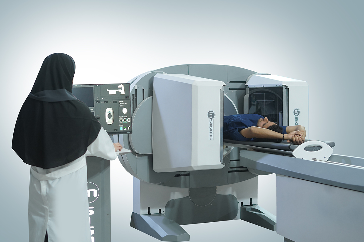
Abstract: A method and a system for single photon emission computed tomography (SPECT) imaging capable of performing a rapid acquisition of imaging data. The SPECT imaging system, placed at fixed radial distance from the center of an object being imaged, includes a gamma detector and a collimator.
Inventors: Hojjat Mahani/Mustafa Abbasi/ Mohammad Reza Ay/ Saeed Sarkar/ Mohammad Hossein Farahani
Abstract:Methods and systems are disclosed, including a method for confining an annihilation range of a positron , from a plurality of positrons emitted from an object being imaged in a positron emission tomography (PET) imaging system. Confining the annihilation includes applying a stochastic multidimensional time varying magnetic field on the positron optionally, the stochastic multidimensional time varying magnetic field includes components in each of three dimensions.
Inventors: Mohammad Reza Ay, Mohammad Hossein Farahani, Saeed Sarkar, Behnoosh Teimourian Fard, Salar Sajedi Toighoun, Sanaz Kaviani
Abstract:
A robotic arm, movable in three rotational degrees of freedom has a base end and a distal end supporting SPECT imaging detectors. A patient support assembly is movable in a linear degree of freedom. A controller causes the robotic arm to move the SPECT imaging detectors, in three dimensions, around the patient.s body to obtain SPECT images. The control causes the patient support assembly to move along the linear degree of freedom, maintaining alignment of the patient.s body with the SPECT imaging detectors.
Inventors: Navid Zeraatkar, Mohammad Hossein Farahani, Mohammad Reza Ay, Saeed Sarkar
Abstract:
An open-gantry structure of SPECT imaging system for scanning human small organs or small animals and method for preparing the system is disclosed. The system contains an imaging desk that one or multiple detector heads are rotated around the object to be scanned while tilted under the imaging desk and dedicated image reconstruction algorithm was developed for the system in case of applying single pinhole collimator.
Inventors: Mohammad Hossein Farahani, Salar Sajedi Toighoun, Mohammad Reza Ay, Saeed Sarkar
Abstract:
A new system and method for medical image processing using a nonlinear recursive filter are disclosed. An input signal including two or more pulses received from a medical imaging system is sampled at a predetermined sampling rate. The maximum magnitude, i.e., peak, and/or the occurrence time of the maximum magnitude of the first pulse of the input signal is/are determined using a nonlinear recursive filter. Predicted magnitude values of the tail of the first pulse can be determined and subtracted from the input signal to correct for pileup before determining the maximum magnitude and/or occurrence time of the next pulses. A medical image can be reconstructed using the determined maximum magnitudes and/or the occurrence times of the maximum magnitudes of the pulses of the input signal. The nonlinear recursive filter can be implemented using one or more look-up tables.
Inventors: Mohammad Reza Ay, Mohammad Hossein Farahani, Afshin Akbarzadeh, Behnoosh Teimourian Fard, Salar Sajedi Toighoun, Navid Zeraatkar
Abstract:
Applications in imaging and spectroscopy rely on pulse processing methods for appropriate data generation. Often, the particular method utilized does not highly impact data quality, whereas in some scenarios, such as in the presence of high count rates or high frequency pulses, this issue merits extra consideration.
In the present study, a new approach for pulse processing in nuclear medicine imaging and spectroscopy is introduced and evaluated. The new non-linear recursive filter (NLRF) performs nonlinear processing of the input signal and extracts the main pulse characteristics, having the powerful ability to recover pulses that would ordinarily result in pulse pile-up. The filter design defines sampling frequencies lower than the Nyquist frequency.
In the literature, for systems involving NaI(Tl) detectors and photomultiplier tubes (PMTs), with a signal bandwidth considered as 15 MHz, the sampling frequency should be at least 30 MHz (the Nyquist rate), whereas in the present work, a sampling rate of 3.3 MHz was shown to yield very promising results. This was obtained by exploiting the known shape feature instead of utilizing a general sampling algorithm. The simulation and experimental results show that the proposed filter enhances count rates in spectroscopy. With this filter, the system behaves almost identically as a general pulse detection system with a dead time considerably reduced to the new sampling time (300 ns). Furthermore, because of its unique feature for determining exact event times, the method could prove very useful in time-of-flight PET imaging.


Lorem ipsum dolor sit amet, consectetur adipiscing elit, sed do eiusmod tempor incididunt ut labore et dolore magna aliqua. Ut enim ad minim veniam, quis nostrud exercitation ullamco laboris nisi ut aliquip ex ea commodo consequat. Duis aute irure dolor in reprehenderit in voluptate velit esse cillum dolore eu fugiat nulla pariatur. Excepteur sint occaecat cupidatat non proident, sunt in culpa qui officia deserunt mollit anim id est laborum.
Devamını oku